2. 730030 兰州,甘肃省骨关节疾病研究重点实验室 ;
3. 730030 兰州, 兰州大学第二医院:血液科
2. Gansu Provincial Key Laboratory of Joint Diseases, Lanzhou, Gansu Province, 730030, China ;
3. Department of Hematology, Lanzhou University Second Hospital, Lanzhou, Gansu Province, 730030
膝关节骨性关节炎(osteoarthritis, OA)和前交叉韧带(anterior cruciate ligament, ACL)损伤是膝关节好发的两大疾病,研究显示OA患者由于骨赘增生会导致髁间窝狭窄[1-2],而髁间窝狭窄又是ACL损伤的危险因素[3]。为了评价髁间窝狭窄,Anderson等[4]和Souryal等[5]先后提出髁间窝宽度指数(notch width index, NWI)的概念,NWI=髁间窝宽度/股骨髁宽度,为相对指标,能降低测量误差,平衡个体差异,较准确的表示髁间窝大小。之后,很多学者对髁间窝不同位置、不同层面的NWI进行了测量,其中Stein等[6]研究OA患者的髁间窝MRI图像时选取了轴位和冠状位两个图层,Hoteya等[7]研究ACL损伤患者的MRI图像时引入了ACL股骨止点的图层,Al-Saeed等[8]提出了MRI图像下髁间窝形状分型的参考标准。但是,到目前为止,对OA患者的髁间窝狭窄与ACL损伤之间关系的研究较少,人们并没有制定出易导致ACL损伤的OA患者MRI图像下不同层面髁间窝狭窄的界值。因此,本研究以中重度OA患者为研究对象,依据是否发生ACL损伤分为两组,分别测量MRI图像下轴位、冠状位、ACL股骨止点位3个层面的NWI,确定OA患者的髁间窝狭窄与ACL损伤之间的关系。
1 资料与方法 1.1 研究对象我们对2011-2014年在兰州大学第二医院拍摄膝关节MRI的1 107例门诊和住院患者进行回顾性病例对照研究。纳入标准:①年龄≥45岁,性别不限;②有膝关节疼痛、晨僵、活动受限、弹响、摩擦感等明确的OA临床表现,符合OA诊断标准[9],X线图像K-L评分[10-11]为Ⅱ、Ⅲ、Ⅳ级的中重度OA患者;③无明确的膝关节外伤或手术史;④MRI图像清晰,序列完整。排除罹患ACL损伤早于OA的患者。所筛选出符合条件的患者的一般资料见表 1。对筛选出的中重度OA患者根据是否并发ACL损伤分为2组,ACL损伤的诊断依据临床表现[12]和MRI征象[13]的相关指南。取得所有患者知情同意,并在医院伦理委员会的监督下进行试验。
| 组别 | 例数 | 年龄 |
| 单纯OA组 | 40 | 56.70±9.14 |
| 男性 | 17 | 55.12±8.71 |
| 女性 | 23 | 57.87±9.46 |
| OA+ACL损伤组 | 35 | 58.03±8.47 |
| 男性 | 8 | 60.13±7.22 |
| 女性 | 27 | 57.41±8.83 |
1.2 测量方法
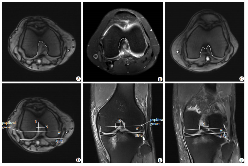
采用Phillips 3.0-T MRI扫描仪,永磁型,配备8通道SENSE膝关节专用线圈。患者仰卧位,膝关节屈曲10°~15°,线圈中心位于髌骨下极水平。扫面序列:轴位质子压脂快速自旋回波(pd-tse-fs-tra)序列,TR 2 886 ms、TE 25 ms,FOV 160 mm,层厚3 mm;冠状位SE序列T1WI,TR 520 ms、TE 20 ms、FOV 160 mm,层厚3 mm。选取序列中显示髁间窝清楚并符合测量位置的图层,用RadiAnt DICOM Viewer 1.9软件进行测量,分型参照Al-Saeed等[8]的标准,分为A、U、W三型(图 1A、B、C)。轴位NWI-1的测量参照Stein等[6]的标准(图 1D),冠状位NWI-2测量参照Domzalski等[3]的标准(图 1E),冠状位NWI-A的测量参照Hoteya等[7]的标准(图 1F)。由3人独立完成,数据取平均值,分型不一致时协商解决。

|
| A:A型,顶端较尖,开口较小,似字母“A”;B:U型,顶端较钝,开口较大,似反向字母“U”;C:W型,顶端双顶,开口较大,似反向字母“W”;D:轴位NWI-1的测量,图层在轴位显示髁间窝最清楚,基线为股骨内外髁软骨下缘的切线,髁间窝深度(D)为髁间窝顶至基线的距离,股骨髁宽度(W)为过股骨外侧髁腘窝沟(popliteal groove)与基线平行的线,髁间窝宽度(N)为D线中上2/3处与基线平行的线;E:冠状位NWI-2的测量,图层在冠状位能看到两条交叉韧带和髁间嵴,基线、W线与图D一致,N线为W线上的髁间窝宽度;F:冠状位NWI-A的测量,图层为冠状位ACL股骨附着点处的图像,W线为过髁间窝出口与基线平行的线,N线为W线上的髁间窝宽度 图 1 膝关节骨性关节炎患者髁间窝形状分型和不同层面NWI的测量 |
1.3 统计学分析
采用SPSS 22.0统计软件,结果以±s的形式表示。不同层面、不同分组间NWI的比较采用单因素方差分析,率的比较采用χ2检验,相关性检验采用Spearman非参数检验。
2 结果 2.1 各组患者一般资料筛选出符合条件的单纯OA组40例,OA合并ACL损伤组35例。并分别通过单因素方差分析及χ2检验得出两组间年龄和性别差异均无统计学意义(P>0.05,表 1),两组之间具有可比性。
2.2 各组患者间髁间窝分型单纯OA组W型1例,OA合并ACL损伤组无W型,因W型较少,且开口宽度与U型接近,所以把W型合并入U型。A型在OA合并ACL损伤组中占88.6%,明显高于单纯OA组(65.0%)。OA合并ACL损伤组与单纯OA组相比分型差异有统计学意义(P < 0.05,表 2)。在中重度OA患者中,A型髁间窝的患者发生ACL损伤的概率是U型的4.173倍(OR=4.173)。
| 组别 | n | A型 | U型 |
| 单纯OA组 | 40 | 26(65.0) | 14(35.0) |
| OA+ACL损伤组 | 35 | 31(88.6)a | 4(11.4)a |
| a:P < 0.05,与单纯OA组比较 | |||
2.3 两组患者间不同层面NWI
与单纯OA组相比,OA合并ACL损伤组不同层面NWI-1、NWI-2、NWI-A均较小(P < 0.01,表 3)。
| 层面 | 单纯OA组 | OA+ACL损伤组 | |||
| x±s | 95%CI | x±s | 95%CI | ||
| NWI-1 | 0.270±0.010 | (0.267~0.274) | 0.257±0.011a | (0.254~0.261) | |
| NWI-2 | 0.252±0.012 | (0.248~0.256) | 0.237±0.017a | (0.231~0.243) | |
| NWI-A | 0.262±0.016 | (0.257~0.267) | 0.240±0.018a | (0.234~0.246) | |
| a:P < 0.01,与单纯OA组比较 | |||||
2.4 两组患者ROC曲线分析
由单纯OA组与OA合并ACL损伤组得到不同层面NWI-1、NWI-2、NWI-A的ROC曲线,曲线下面积分别为0.805、0.754、0.808(P均<0.01),最佳界值为约登指数(灵敏度+特异度-1)最大点对应的值,得出最佳界值分别为0.263、0.248、0.254。NWI-A的ROC曲线下面积最大(图 2)。以上界值均小于等于单纯OA组对应平面NWI的95%可信区间下限,且ROC曲线下面积大于0.5(P < 0.01),因此,以上界值是合理的。

|
| A、B、C分别为NWI-1、NWI-2、NWI-A的ROC曲线 图 2 各组患者不同层面NWI的ROC曲线分析 |
2.5 各组患者不同层面NWI的灵敏度和特异度
以小于ROC曲线最佳界值为中重度OA患者髁间窝严重狭窄的依据,即NWI-1<0.263、NWI-2<0.248、NWI-A<0.254为OA患者髁间窝严重狭窄,反之为非严重狭窄。得出:NWI-1<0.263在诊断OA患者合并ACL损伤的灵敏度是71.4%,特异度是80.0%;NWI-2<0.248在诊断OA患者合并ACL损伤的灵敏度是71.4%,特异度是67.5%;NWI-A<0.254在诊断OA患者合并ACL损伤的灵敏度是80.0%,特异度是70.0%。NWI-A<0.254在诊断OA患者合并ACL损伤的灵敏度最高,NWI-1<0.263的特异度最高(表 4)。
| 组别 | n | NWI-1 | NWI-2 | NWI-A |
| OA+ACL损伤组 | 35 | |||
| 严重狭窄 | 25(71.4) | 25(71.4) | 28(80.0) | |
| 非严重狭窄 | 10(28.6) | 10(28.6) | 7(20.0) | |
| 单纯OA组 | 40 | |||
| 严重狭窄 | 8(20.0) | 13(32.5) | 12(30.0) | |
| 非严重狭窄 | 32(80.0) | 27(67.5) | 28(70.0) |
3 讨论
膝关节OA是一种退行性疾病,由于关节软骨炎症、退化,软骨下骨的反应性增生,而形成骨赘。众多学者对OA患者的髁间窝进行了研究,Shepstone等[14]发现,健康人髁间窝内侧壁向内凹陷,而OA患者内侧壁比较直甚至向外凸出,这种形态差异可能会减小髁间窝宽度。Wada等[1]在32例全膝关节置换的患者和54具尸体标本上对髁间窝行解剖学研究,发现OA患者髁间窝内由于骨赘的增生可出现髁间窝狭窄,OA越严重,骨赘越多,髁间窝狭窄程度越重,分级为Ⅲ、Ⅳ级的OA明显比Ⅰ级的患者NWI小很多。León等[2]发现OA患者可出现各种类型的骨赘增生,OA越严重增生越明显,这种增生必然会减小髁间窝宽度,形成髁间窝狭窄,导致NWI减小。
关于髁间窝形状,多数学者把髁间窝分成了A、U、W三型。Anderson等[4]对CT图像研究发现U型的髁间窝不易发生狭窄,van-Eck等[15]在关节镜下发现A型的髁间窝宽度整体上比U型小。Sutton等[16]发现髁间窝分型与性别有关,女性的髁间窝宽度比男性小,且A型较多,说明A型髁间窝容易狭窄。Al-Saeed等[8]研究MRI图像时发现A型髁间窝较窄,且髁间窝形状是ACL损伤的一个危险因素,A型易发生ACL损伤。本研究发现OA合并ACL损伤患者A型所占比例明显高于单纯OA患者,说明在OA患者中,A型的髁间窝也容易出现ACL损伤。
近年来ACL损伤的发病率逐渐升高,在运动员及军人中尤为常见[17]。大量研究证实髁间窝狭窄是ACL损伤的独立危险因素,ACL损伤患者的NWI比健康人小[3, 18-22],且左右双侧ACL损伤的患者NWI比单侧损伤的患者更小[5, 7]。其中Hoteya等[7]选取了ACL股骨止点层面的NWI-A与其后层面的NWI-P进行测量,发现ACL损伤组的NWI-A、NWI-P均比健康人小,但我们认为NWI-P层面无ACL走形,因此对ACL损伤的指导意义不大,所以只选取了NWI-A层面。对于髁间窝狭窄导致ACL损伤的原因,可能与髁间窝狭窄患者的ACL横截面积较小、强度较弱,生物学性能较差,易发生损伤有关[23-24];也可能与在膝关节运动时ACL与狭窄的髁间窝发生碰撞,产生磨损,导致ACL损伤有关[25]。León等[2]发现OA患者可出现各种类型的骨赘增生,导致不同部位的髁间窝撞击,当髁间窝顶增生时可出现ACL前部撞击,当髁间窝出口处增生时可出现ACL中部撞击,当顶部和出口都增生时,ACL前部和中部都会出现撞击,而髁间窝严重狭窄的Ⅳ级OA,髁间窝内大部增生,会出现ACL大部的挤压变形,这种长时间的髁间窝撞击会造成ACL不可逆性损伤。X线片和CT图像研究发现,在OA合并ACL损伤患者的髁间窝宽度、角度和面积均比单纯OA患者小,OA合并ACL断裂缺失的患者的以上指标更小[26]。解剖学研究发现OA患者中可出现大量的ACL损伤,且ACL损伤程度越重,NWI越小[1]。分子生物学研究发现狭窄的髁间窝磨损ACL,可使磨损区域的金属基质蛋白酶活性增高,胶原及硫酸化糖胺聚糖降解增加,从而使ACL造成不可逆性破坏[27]。Stein等[6]对160例不同严重程度的OA患者行MRI评估发现,合并ACL损伤患者的轴位和冠状位NWI均明显小于单纯OA患者。也有大量研究发现年轻人ACL损伤,最终会导致OA的形成,主要原因是ACL损伤后关节不稳,进而改变局部生物力学,会破坏关节软骨,产生关节炎症,导致骨赘增生,骨赘增生后会进一步加重关节磨损,最终导致OA[28-29]。即使在ACL损伤后进行重建仍不能有效阻止OA的发生[30-31]。OA患者由于骨赘的增生,引起髁间窝狭窄,可以诱发ACL损伤,而ACL损伤后由于局部生物力学改变导致的软骨退变又可加重OA,鉴于ACL损伤与OA的这种相互关系,对髁间窝狭窄的OA患者提前采取预防、治疗措施,对防止ACL损伤并发症的出现及缓解OA病程的进展至关重要。
对不同层面NWI髁间窝狭窄界值的确定,Anderson等[4]提出以X线片NWI<0.20为依据,Hoteya等[7]提出以MRI图像冠状位ACL股骨止点处NWI-A<0.25为依据,但作者均未提出界值具体的制定标准,且关于OA患者髁间窝狭窄的研究较少,尚未提出明确界值。本研究以ROC曲线最佳界值为标准,确定了OA患者轴位NWI-1<0.263、冠状位NWI-2<0.248、ACL股骨止点位NWI-A<0.254的髁间窝狭窄界值,为OA患者髁间窝狭窄的确定提供了参考依据。对髁间窝狭窄的OA患者采取定时体检、及时提供防治建议、减轻体质量、减少关节负重、限制运动、应用软骨保护剂(如关节腔内注射玻璃酸钠或口服硫酸氨基葡萄糖)等措施有助于ACL损伤的预防。
综上所述,OA患者的ACL损伤与其髁间窝狭窄程度相关,合并ACL损伤的OA患者明显比单纯OA患者髁间窝狭窄,且A型较多。结合我们之前的研究可以得出结论:髁间窝狭窄可能是OA患者发生ACL损伤的危险因素[32-34]。对髁间窝不同层面NWI小于界值的髁间窝狭窄患者及时提供防治措施对OA患者ACL损伤的发生起到一定的预防作用。但由于本研究病例仅来自于一家医院,取材范围较窄,样本量不够大,因此,相关结论尤其是界值的确定,需要多中心、大样本研究进一步证实。
| [1] | Wada M, Tatsuo H, Baba H, et al. Femoral intercondylar notch measurements in osteoarthritic knees[J]. Rheumatology (Oxford),1999, 38 (6) : 554 –558. DOI:10.1093/rheumatology/38.6.554 |
| [2] | León H O, Blanco C E, Guthrie T B, et al. Intercondylar notch stenosis in degenerative arthritis of the knee[J]. Arthroscopy,2005, 21 (3) : 294 –302. DOI:10.1016/j.arthro.2004.11.019 |
| [3] | Domzalski M, Grzelak P, Gabos P. Risk factors for Anterior Cruciate Ligament injury in skeletally immature patients: analysis of intercondylar notch width using Magnetic Resonance Imaging[J]. Int Orthop,2010, 34 (5) : 703 –707. DOI:10.1007/s00264-010-0987-7 |
| [4] | Anderson A F, Lipscomb A B, Liudahl K J, et al. Analysis of the intercondylar notch by computed tomography[J]. Am J Sports Med,1987, 15 (6) : 547 –552. DOI:10.1177/036354658701500605 |
| [5] | Souryal T O, Moore H A, Evans J P. Bilaterality in anterior cruciate ligament injuries: associated intercondylar notch stenosis[J]. Am J Sports Med,1988, 16 (5) : 449 –454. DOI:10.1177/036354658801600504 |
| [6] | Stein V, Li L, Guermazi A, et al. The relation of femoral notch stenosis to ACL tears in persons with knee osteoarthritis[J]. Osteoarthritis Cartilage,2010, 18 (2) : 192 –199. DOI:10.1016/j.joca.2009.09.006 |
| [7] | Hoteya K, Kato Y, Motojima S, et al. Association between intercondylar notch narrowing and bilateral anterior cruciate ligament injuries in athletes[J]. Arch Orthop Trauma Surg,2011, 131 (3) : 371 –376. DOI:10.1007/s00402-010-1254-5 |
| [8] | Al-Saeed O, Brown M, Athyal R, et al. Association of femoral intercondylar notch morphology, width index and the risk of anterior cruciate ligament injury[J]. Knee Surg Sports Traumatol Arthrosc,2013, 21 (3) : 678 –682. DOI:10.1007/s00167-012-2038-y |
| [9] | 中华医学会风湿病学分会. 骨关节炎诊断及治疗指南[J]. 中华风湿病学杂志,2010, 14 (6) : 416 –419. DOI:10.3760/cma.j.issn.1007-7480.2010.06.024 |
| [10] | Kellgren J H, Lawrence J S. Radiological assessment of osteo-arthrosis[J]. Ann Rheum Dis,1957, 16 (4) : 494 –502. DOI:10.1136/ard.16.4.494 |
| [11] | Kim H J, Lee J Y, Kim T J, et al. Association between serum vitamin D status and health-related quality of life (HRQOL) in an older Korean population with radiographic knee osteoarthritis: data from the Korean national health and nutrition examination survey (2010-2011)[J]. Health Qual Life Outcomes,2015, 13 : 48 . DOI:10.1186/s12955-015-0245-1 |
| [12] | 中华中医药学会. 膝关节交叉韧带损伤[J]. 风湿病与关节炎,2013, 2 (5) : 78 –80. |
| [13] | 廖云, 孙仁权, 廖国栋. MRI多征象分析在膝关节前交叉韧带损伤诊断中的临床应用价值分析[J]. 中国CT和MRI杂志,2015, 13 (6) : 101 –103. DOI:10.3969/j.issn.1672-5131.2015.06.032 |
| [14] | Shepstone L, Rogers J, Kirwan J R, et al. Shape of the intercondylar notch of the human femur: a comparison of osteoarthritic and non-osteoarthritic bones from a skeletal sample[J]. Ann Rheum Dis,2001, 60 (10) : 968 –973. DOI:10.1136/ard.60.10.968 |
| [15] | van-Eck C F, Martins C A, Vyas S M, et al. Femoral intercondylar notch shape and dimensions in ACL-injured patients[J]. Knee Surg Sports Traumatol Arthrosc,2010, 18 (9) : 1257 –1262. DOI:10.1007/s00167-010-1135-z |
| [16] | Sutton K M, Bullock J M. Anterior cruciate ligament rupture: differences between males and females[J]. J Am Acad Orthop Surg,2013, 21 (1) : 41 –50. DOI:10.5435/JAAOS-21-01-41 |
| [17] | 谢峰, 吕慈, 黄昌林, 等. 陆地军训所致膝关节前交叉韧带损伤的伤情特点及诊疗效果分析[J]. 第三军医大学学报,2015, 37 (13) : 1373 –1376. DOI:10.16016/j.1000-5404.201411031 |
| [18] | Keays S L, Keays R, Newcombe P A. Femoral intercondylar notch width size: a comparison between siblings with and without anterior cruciate ligament injuries[J]. Knee Surg Sports Traumatol Arthrosc,2016, 24 (3) : 672 –679. DOI:10.1007/s00167-014-3491-6 |
| [19] | Sonnery-Cottet B, Archbold P, Cucurulo T, et al. The influence of the tibial slope and the size of the intercondylar notch on rupture of the anterior cruciate ligament[J]. J Bone Joint Surg Br,2011, 93 (11) : 1475 –1478. DOI:10.1302/0301-620X.93B11.26905 |
| [20] | Park J S, Nam D C, Kim D H, et al. Measurement of Knee Morphometrics Using MRI: A Comparative Study between ACL-Injured and Non-Injured Knees[J]. Knee Surg Relat Res,2012, 24 (3) : 180 –185. DOI:10.5792/ksrr.2012.24.3.180 |
| [21] | Ireland M L, Ballantyne B T, Little K, et al. A radiographic analysis of the relationship between the size and shape of the intercondylar notch and anterior cruciate ligament injury[J]. Knee Surg Sports Traumatol Arthrosc,2001, 9 (4) : 200 –205. DOI:10.1007/s001670100197 |
| [22] | Souryal T O, Freeman T R. Intercondylar notch size and anterior cruciate ligament injuries in athletes. A prospective study[J]. Am J Sports Med,1993, 21 (4) : 535 –539. DOI:10.1177/036354659302100410 |
| [23] | Stijak L, Bumbasirevic M, Kadija M, et al. Morphometric parameters as risk factors for anterior cruciate ligament injuries-a MRI case-control study[J]. Vojnosanit Pregl,2014, 71 (3) : 271 –276. DOI:10.2298/VSP1403271S |
| [24] | Dienst M, Schneider G, Altmeyer K, et al. Correlation of intercondylar notch cross sections to the ACL size: a high resolution MR tomographic in vivo analysis[J]. Arch Orthop Trauma Surg,2007, 127 (4) : 253 –260. DOI:10.1007/s00402-006-0177-7 |
| [25] | Everhart J S, Flanigan D C, Simon R A, et al. Association of noncontact anterior cruciate ligament injury with presence and thickness of a bony ridge on the anteromedial aspect of the femoral intercondylar notch[J]. Am J Sports Med,2010, 38 (8) : 1667 –1673. DOI:10.1177/0363546510367424 |
| [26] | Hernigou P, Garabedian J M. Intercondylar notch width and the risk for anterior cruciate ligament rupture in the osteoarthritic knee: evaluation by plain radiography and CT scan[J]. Knee,2002, 9 (4) : 313 –316. DOI:10.1016/S0968-0160(02)00053-4 |
| [27] | Comerford E J, Tarlton J F, Avery N C, et al. Distal femoral intercondylar notch dimensions and their relationship to composition and metabolism of the canine anterior cruciate ligament[J]. Osteoarthritis Cartilage,2006, 14 (3) : 273 –278. DOI:10.1016/j.joca.2005.09.001 |
| [28] | Simon D, Mascarenhas R, Saltzman B M, et al. The Relationship between Anterior Cruciate Ligament Injury and Osteoarthritis of the Knee[J]. Adv Orthop,2015, 2015 : 928301 . DOI:10.1155/2015/928301 |
| [29] | Svoboda S J. ACL injury and posttraumatic osteoarthritis[J]. Clin Sports Med,2014, 33 (4) : 633 –640. DOI:10.1016/j.csm.2014.06.008 |
| [30] | Janssen R P, du-Mee A W, van-Valkenburg J V, et al. Anterior cruciate ligament reconstruction with 4-strand hamstring autograft and accelerated rehabilitation: a 10-year prospective study on clinical results, knee osteoarthritis and its predictors[J]. Knee Surgery Sports Traumatol Arthrosc,2013, 21 (9) : 1977 –1988. DOI:10.1007/s00167-012-2234-9 |
| [31] | Barenius B, Ponzer S, Shalabi A, et al. Increased risk of osteoarthritis after anterior cruciate ligament reconstruction: a 14-year follow-up study of a randomized controlled trial[J]. Am J Sports Med,2014, 42 (5) : 1049 –1057. DOI:10.1177/0363546514526139 |
| [32] | Chen C, Ma Y, Geng B, et al. Intercondylar Notch Stenosis of Knee Osteoarthritis and Relationship between Stenosis and Osteoarthritis Complicated with Anterior Cruciate Ligament Injury: A Study in MRI[J]. Medicine (Baltimore),2016, 95 (17) : e3439 . DOI:10.1097/MD.0000000000003439 |
| [33] | 陈聪, 马愔花, 谭小义, 等. 骨性关节炎与髁间窝狭窄之间关系的研究[J]. 中国矫形外科杂志,2016, 24 (1) : 73 –78. DOI:10.3977/j.issn.1005-8478.2016.01.15 |
| [34] | 陈聪, 马愔花, 谭小义, 等. 冠状位髁间窝宽度指数与骨关节炎的关系[J]. 南方医科大学学报,2015, 35 (10) : 1384 –1389. DOI:10.3969/j.issn.1673-4254.2015.10.04 |


